|
Advanced Navigator Techniques
K. Nehrke and D. Manke*
Philips Research Laboratories, Division Technical Systems,
Röntgenstrasse 24-26, D-22335 Hamburg, Germany
* Institute of Biomedical Engineering, Universität Karlsruhe,
Kaiserstrasse 12, D-76128 Karlsruhe, Germany
Correspondence: kay.nehrke@philips.com
Abstract. The purpose of this study was to investigate and to optimize the performance
of the real-time navigator technology on a clinical scanner for use in
cardiac imaging. The studies involved experiments performed on phantoms
and in vivo. The performance of the 2D RF pulses used for pencil beam excitation
was found to be highly sufficient, provided a proper pulse design, accurately
trimmed gradients and reasonable pulse parameters were used. The capability
of the navigators for free-breathing, real-time gated coronary artery imaging
was studied for several volunteers. Using a diaphragmatic gating window
of 5 mm a reproducible image quality was achieved. To study spatial and
temporal correlations of respiratory motion multiple navigator pulses were
applied in different anatomical regions. For the correlation diaphragm-heart
strong deviations from the linear model reported in literature were found.
The results indicate, that a more complex model, including patient dependent
hysteretic effects, might further improve the performance of real-time
gating and prospective motion correction.
Keywords: Navigator, coronary artery
imaging, real-time gating, 2D RF pulses
Introduction
Respiratory motion can severely deteriorate
the image quality of long cardiac-triggered MR imaging sequences. Therefore,
gating based on navigator echoes [1,2]
was introduced to reduce these artefacts. By means of a navigator, the
position of the diaphragm can be monitored [3] and
used as an input for the accept/reject decision of the gating algorithm.
Furthermore, the navigator information may be used to perform prospective
motion correction like slice-tracking to improve image quality and/or to
allow a larger gating window [4]. On the other hand,
robust real-time gating makes several demands on the performance of the
navigator technique. The displacement of a selected anatomical region has
to be monitored with high accuracy and destructive interference of the
navigator pulse with the imaging volume has to be avoided. The navigator
sequence should be short enough to allow versatile integration into MR
imaging protocols. In addition, a fast evaluation of the measured displacements
is necessary for real-time gating. Furthermore, an appropriate model
for the respiratory motion of the heart is required, if prospective motion
correction is performed. The purpose of this study was to investigate and
to optimize the performance of a navigator on a clinical scanner with respect
to these demands.
Methods
In vivo experiments with several healthy volunteers
and phantom experiments were performed on a 1.5 T whole body scanner (GYROSCAN
ACS NT, Philips Medical Systems) with self-shielded gradients (23 mT in
0.2 ms).
Navigator pulse design
For the navigator 2D RF pulses are used [5].
These excite a spatially restricted volume of pencil beam shape, which
is read out using a gradient echo. This allows to monitor in-vivo motion
along one direction (fig.1). The 2D RF pulses are based on a spiral
k-space trajectory (fig. 2), played out by the gradient system in the presence
of a B1-field. To attain optimal results, the gradient
performance, a proper pulse design and the aliasing problem have
to be considered. The quality of the gradient system employed affects the
performance and positioning accuracy of a navigator pulse. Due to the discrete
k-space covering of the spiral trajectory aliasing rings are excited, which
degrade the 1D navigator profiles. To shift the aliasing rings out of the
body, the number of k-space turns has to be increased. Due to gradient
constraints this is accompanied by an increase of the pulse duration
and, hence, of the off-resonance sensitivity of the 2D RF pulse. It is
always necessary to find an appropriate compromise between the pencil beam
diameter, the aliasing and the off-resonance sensitivity [6].
To investigate the spatial distribution of magnetization of the pencil
beam, the 2D RF-pulse was used for excitation in an imaging sequence. Phantom
and in-vivo experiments were performed to study the positioning accuracy,
the spatial selectivity and the aliasing of the 2D RF pulse.

Figure 1. Coronal survey with pencil beam through diaphragm
(left). The Fourier transform of the gradient echo yields the projection
of the pencil beam's magnetization onto the z-axis, the so-called navigator
profile, which is sketched on the right for two different diaphragm positions.
The contrast change between liver and lung results in a step-like shape.
The displacement of the step with respect to a reference profile is determined
in real-time by a cross-correlation algorithm. The total duration of the
navigator pulse sequence including exciation, acquisition and evaluation
is currently 20 ms.
Figure 2. Spiral trajectory of 2D-RF pulse (left) and
corresponding point spread function (right). The aliasing rings appear
due to the discrete k-space covering of the spiral. The central peak is
used as a pencil beam navigator.
Real-time gating
The capability of the navigators for real-time
respiratory gating was examined in free-breathing coronary MR angiography,
where the pencil beam was applied through the right hemidiaphragm. The
proximal portions of the right coronary arteries (RCA) and the left anterior
descending artery (LAD) were imaged using an ECG-triggered segmented k-space
3D gradient echo sequence (TR=8.6 ms, TE = 3.2 ms, flip-angle = 30°,
512x358 pixel matrix). For respiratory gating an acceptance window of 5
mm was chosen.
Multiple navigators
To study the respiratory motion of the heart
during free breathing multiple navigator pulses were used. The scan software
was extended to provide up to four independent navigator pulses, which
can be positioned and angulated freely in space. The respiratory motion
of right hemidiaphragm, left ventricle, chest wall and abdominal wall was
recorded over 10 minutes, using a pure navigator sequence with high temporal
resolution (20 ms per navigator). The correlation between the different
navigators was analyzed in 2D-histograms.
Results
Navigator pulse performance
The navigator performance was found to be highly
sufficient for most applications. Using a simple eddy-current precompensation
scheme for the gradient waveforms to correct for residual short-term eddy
currents, an accuracy of positioning of the pencil beam better than 7 mm
was achieved, which is only a fraction of a typical pencil beam diameter
of 25 mm. Using strong gradient slew-rates (100 mT/m/ms) it was always
possible to shift the aliasing rings out of the body without inducing off-resonance
problems due to the longer pulse length (fig. 3). The spatial selectivity
of the 2D-RF pulse was improved by proper weighting of the RF waveform.
The navigator profiles obtained from the navigator echo show a well defined
change in contrast at the lung-tissue interface (fig. 4). Hence, the displacement
of the navigator profiles could be determined reliably by the cross-correlation
algorithm. By sub-pixel interpolation an accuracy better than 1mm was achieved.

Figure 3. Transversal spin-echo images of the pencil beam excitation
in the abdominal region (b-d). In the transversal survey (a) the expected
positions of the central beam and the aliasing rings for a 3-turn spiral
are indicated by white circles. With increasing number of k-space turns
(b-d: 3,6,12) the rings are shifted out of the body. The corresponding
pulse lengths are 1.7ms, 2.8 ms, and 5.7 ms, respectively.

Figure 4. Coronal survey indicating navigator positions (left),
and corresponding navigator profiles (right). The red points indicate the
respiration curves determined by cross-correlation in real-time. For the
navigator through the heart a contribution from cardiac motion is superimposed.
Coronary artery imaging
In figure 5 a vessel-based maximum intensity
projection of the RCA and the LAD is shown for one selected volunteer.
The branching out of the vessels is well resolved, indicating the efficacy
of the gating approach. In the volunteer experiments the gating efficiency
was between 20% and 60 %, dependent on the individual motion pattern of
the subject. For the applied imaging protocol this corresponds to scan
times between 10 and 30 minutes.
Figure 5. RCA (left) and LAD (right) of a healthy volunteer.
The in-plane resolution of the 3D data set was 0.7 mm, the through-plane
resolution 1.5 mm.
Respiratory motion of the heart
The 2D histograms in fig. 6 show the correlation
between the diaphragmatic motion and the superior-inferior motion of the
left ventricle for four selected volunteers. For all volunteers, well defined
trajectories without much scattering were found. This indicates a good
correlation between diaphragm and heart. However, the histograms show also
hysteresis loops with different branches for expiration and inspiration.
Hysteresis means, that a certain diaphragm position corresponds to different
heart positions for inspiration and expiration, respectively. This hysteretic
behavior is strongly patient dependent, for some volunteers a perfect linear
relationship is observed, for others a large gap up to 6 mm with respect
to the heart position was found. These results indicate, that a simple
linear mapping of the diaphragmatic motion onto the respiratory motion
of the heart as suggested in the literature
[7],
may lead to considerable errors in case of tracking the imaging slab. Instead,
a patient dependent calibration of the correlation diaphragm-heart should
be performed prior to the MR imaging scan by using multiple navigators.
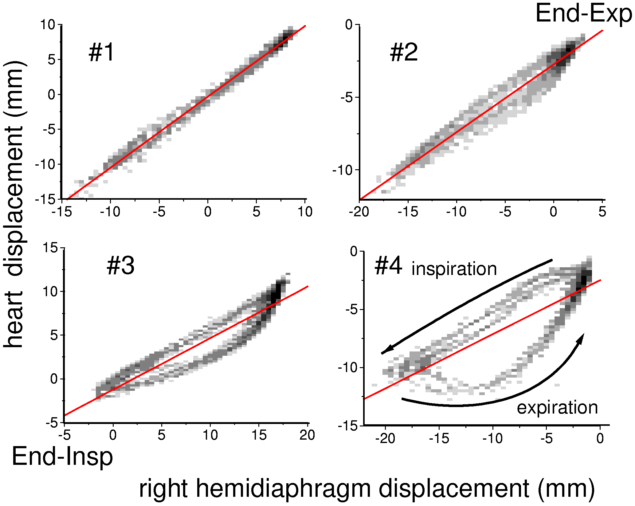
Figure 6. 2D histogram plots of the correlation heart-diaphragm
for four selected volunteers. The corresponding navigator positions are
shown in fig.4. Black bins indicate many counts, light bins indicate few
counts. The red lines are linear fits to the data.
Conclusions
Pencil beam navigator pulses represent a powerful
approach to monitor in vivo motion. When used for real-time gating high
resolution coronary artery images can be obtained with reproducible quality
during free breathing. The multiple navigator results indicate, that a
proper model for the correlation diaphragm-heart, including patient dependent
hysteretic effects, might further improve image quality or decrease scan
time, when used for prospective motion correction. Multiple navigator pulses
offer the potential for an automatic, patient dependent calibration of
such a model.
References
[1] Liu, Y.L., Riederer,
S.J., Rossmann, P.J., Grimm, R.C., Debbins, J.P. and Ehman, R.L., "A monitoring,
feedback, and triggering system for reproducible breath-hold MR imaging",
Magnetic Resonance in Medicine, vol. 30, p. 507, 1993.
[2] Sachs, T.S., Meyer,
C.H., Hu, B.S., Kohli, J., Nishimura, D. and Macovski, A., "Real-time motion
detection in spiral MRI using navigators", Magnetic Resonance in Medicine,
vol. 32, p. 639, 1994.
[3] Wang, Y., Grimm,
R.C., Felmlee, J.P., Riederer, S.J. and Ehman, R.L., "Algorithms for extracting
motion information from navigator echoes", Magnetic Resonance in Medicine,
vol. 36, p. 117, 1996.
[4] Ehman, R.L. and
Felmlee, J.P., "Adaptive technique for high-definition MR imaging of moving
structures", Radiology, vol. 173, p. 255, 1989.
[5] Pauly, J., Nishimura,
D. and Macovski, A., "A k-space analysis of small-tip-angle excitations",
Magnetic Resonance in Medicine, vol. 81, p. 43, 1989.
[6] Nehrke, K., Börnert,
P., Groen, J., Smink, J. and Böck, J.C, "On the performance and accuracy
of 2D navigator pulses", Magnetic Resonance Imaging, vol. 17, no. 8, p.
1173, 1999.
[7] Wang, Y., Riederer, S.J.
and Ehman, R.L.,"Respiratory motion of the hart: kinematics and the implications
for spatial resolution in coronary imaging", Magnetic Resonance in Medicine,
vol. 33, p. 713, 1995.
|

 Home
Current Issue
Table of Contents
Home
Current Issue
Table of Contents


