|
International Journal of Bioelectromagnetism Vol. 5, No. 1, p. 268, 2003. |
www.ijbem.org |
|
Does Left Atrial Wall Strain Influence
on SA ECG and
A. Glowniak, A. Kutarski, D. Szczesniak, P. Rucinski, and T. Widomska-Czekajska
Department of Cardiology, University Medical Academy of Lublin, Poland Abstract. The time domain signal-averaged
(SA) P wave analysis is a valuable method for detection of atrial conduction
disturbances which may indicate increased risk of atrial arrhythmias. But
the conjunction of presence of positive criteria of atrial late potentials
(prolonged P duration and decreased RMS 20 values) and increased strain of
atrial wall/atrial enlargement remain unclear.
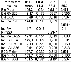
Our goal was to evaluate correlation between conventional USG findings and EPS properties of atria evaluated using high gain SA ECG and SA right and left atrial IEGM. Method. Examinations (during SR) were performed in 24 pts during implantation of biatrial pacing system. External signals were obtained from Frank orthogonal leads. Intra-atrial signals were recorded separately from right and left atrium, using bipolar pacing leads placed in RAA and CS and ventricular lead temporary placed in LRA. Signals were filtered and recorded with Codax SAI-IK amplifier, digitized by A/D converter and stored on PC. We analyzed following SAECG parameters: P wave duration (Pdur), root mean square voltages of the last 20 ms of P wave (RMS20) and duration of low amplitude signal < 5mV (LAS5). We considered ALP as positive if Pdur > 125 ms and RMS20 < 2.40 mV. Results. Conclusion. LVDd and EF correlates with SA P wave duration but values of mentioned parameters seems not to influence upon presence of micro oscillations during final part of atrial excitation.
© International Society for Bioelectromagnetism
|