|
International Journal of Bioelectromagnetism Vol. 4, No. 2, pp. 257-258, 2002. |
www.ijbem.org |
|
Influence of realistic head geometry differences
K. Whittingstall1, G.
Stroink1,4, L. Gates2,J.F. Connolly3, and G.
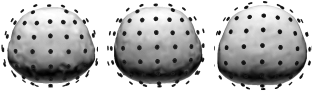
A. Finley4 Abstract: To enhance the signal to noise level in event related potential (ERP) measurements, one frequently resorts to obtaining a grand average of the data obtained across a group of subjects. This averaged data is then used in EEG source localization. Since it is often not feasible to obtain realistic head models for each of the subjects, a sphere or average realistic head is used for source reconstruction. However, this procedure does not account for head geometry differences between subjects, thereby giving rather tentative descriptions of source locations. The present study uses computer simulations of ERP measurements and realistic head shapes constructed from individual MRI scans to evaluate the effect of varying head shape on the accuracy of this dipole localization procedure. Errors in source localization were on average 1.1cm (SD=0.5cm) for SNR values ranging from 4-60 and 2.0cm (SD=1cm) for SNR values ranging from 2-3. INTRODUCTIONElectroencephalograms (EEGs) are often used for the estimation of equivalent current sources that is thought to represent brain activity. Several techniques for the solution of the so-called inverse problem, such as the multiple signal classification (MUSIC) algorithm [1], have been developed. In order to solve the EEG forward problem as part of this inverse solution, a head model is needed. Influences of realistic and spherical head models as volume conductors on source localization [2,3] have been investigated, as has its influence due to electrode misplacement [4]. Due to their relatively low signal to noise ratio (SNR), it is difficult to locate sources using recorded EEGs on an individual basis. Therefore, in an attempt to enhance the SNR of the recorded EEG, one frequently obtains a grand average of the data obtained from a group of subjects. Since it is often not feasible to obtain MRI scans of each individual, this averaged data in conjunction with a sphere or "average" head model is commonly used in EEG source localization techniques [5,6]. Such an approximation will introduce errors in the source modeling procedure. The present study uses MRI constructed head shapes and computer simulations of ERP measurements to evaluate the accuracy of this dipole localization procedure in noisy signal environments. METHODSRealistically shaped 3-layer boundary element head models were constructed from MRI scans of 5 subjects using CURRY software. The conductivity of the brain, skull and scalp were set to 0.33, 0.0042, and 0.33 S/m, respectively. The 10-20 International System was used to project 64 electrodes on the surface scalp using fiducial markers placed on anatomical landmarks prior to image acquisition. Simulated time varying signals from 4 sources located in well-separated areas of the brain from one of the realistically shaped head models were generated at 64 electrode sites. Two sources were located approximately 1 and 4 cm below the scalp in each hemisphere. Six sets of simulated noise were then added to simulate realistic EEG recording conditions. The SNR was estimated by using a signal free interval. The acquired simulated signal was then projected on the other 4 different realistic head models. Dipole localization was carried out using the MUSIC algorithm as implemented in the CURRY software. The resultant dipole locations were compared with the original locations of the sources in the reference model to investigate errors of source localization caused by differences in head geometry at different noise levels. Figure 1 shows segmented skulls with associated electrode montage from 3 of the 5 subjects used in this study. The leftmost skull represents the model from which the simulated data was constructed. This was also used as a reference for all source localization calculations.
Figure 1. Variations in skull geometry between the reference model (far left) and two of the four models used to investigate source localization errors. All head models were placed in the same coordinate system by matching anatomical landmarks between subjects. Table 1 shows the average variation in electrode placement of 4 different subject head models relative to the reference model. TABLE 1
RESULTSThe mean and standard deviation of the dipole source localization errors caused by geometry differences at different SNR levels are shown in Fig. 2. Figure 2. Average localization error of four different sources in four different realistic head models relative to the reference model at different SNR values. As anticipated, the average localization error remains relatively constant at high SNR but rapidly increases at lower SNR. To investigate effects of electrode placement differences on source locations due to different head geometry, the average localization errors for the 4 realistic models were divided into two SNR ranges. Figure 3 shows localization errors due to electrode location differences in two different SNR ranges. Figure 3. Source localization errors due to variation in electrode placement at high SNR (solid line represents standard deviation) and low SNR (dashed line represents standard deviation). The effects of variations in electrode placement appear well behaved at high SNR values, whereas dipole solutions at low SNR are less accurate and less stable. This figure demonstrates that variations in electrode location due to different head geometries are the main cause of the localization errors at high SNR ranges. DISCUSSION Source localization with average subject data combined with a realistic average head model do not take into account differences in actual head geometry and can introduce error in the location of the source(s). Overall, our results show that if grand average waveforms are to be used to determine the location of sources, one can expect localization errors of approximately 1.1cm (SD=0.5cm) for high (4-60) SNR and about 2 cm (SD=1cm) for low (2-3) SNR. It is anticipated that this error increases with even lower SNR values. Variations in electrode placement due to different head geometries appear to have a significant impact on the localization procedure at high SNR ranges. However, this error due to taking averages over a group of subjects may be tolerable when drawing general conclusions about the source location. At low SNR ranges, the noise itself is the dominant factor in localization errors. ACKNOWLEDGEMENTSThis work is supported, in part, by the Natural Science and Engineering Council (NSERC). We also would like to thank Dr. P. McGrath for his encouragement. REFERENCES[1] A. Mosher, P. Lewis, R. Leahy. Multiple Dipole Modeling and Localization from Spatio-Temporal MEG data IEEE Trans. Biomed. Eng., vol. 39, pp. 541-557, 1992. [2] B.N. Cuffin. Effects of Head Shape on EEGs and MEGs IEEE Trans. Biomed. Eng. Vol. 37, pp. 44-52, 1990 [3] B.N. Cuffin. EEG Localization Accuracy Improvements Using Realistically Shaped Head Models IEEE Trans. Biomed. Eng. Vol. 43, pp. 299-303, 1996 [4] Y. Wang, J. Gotman. The influence of electrode location errors on EEG dipole source localization with a realistic head model Clin. J. Neurophys. Vol. 112, pp. 1777-1780, 1990 [5] M. Scherg, D. Von Cramon. Evoked Dipole Source Potentials of the Human Auditory Cortex Electroencephalography and Clinical J. Neurophys vol. 65, pp. 344-360, 1986. [6] B. Opitz, A. Mecklinger, D.Y. Von Cramon and F. Kruggel. Combining electrophysiological and hemodynamic measures of the auditory oddball Physcphysiology, vol. 36, pp.142-147, 1999
© International Society for Bioelectromagnetism
|